
Press images
Pictures may only be used be used in connection with the Microbe of the Year and with reference to the source.
Herunterladen
Press images/ files Microbe of the Year 2025 (Corynebacterium glutamicum)

Morphology of Corynebacterium glutamicum: Transmission electron micrograph with storage substances such as polyphosphate and lipid droplets. Image: Marc Bramkamp and Urska Repnik, Kiel University (CC BY 4.0).
Herunterladen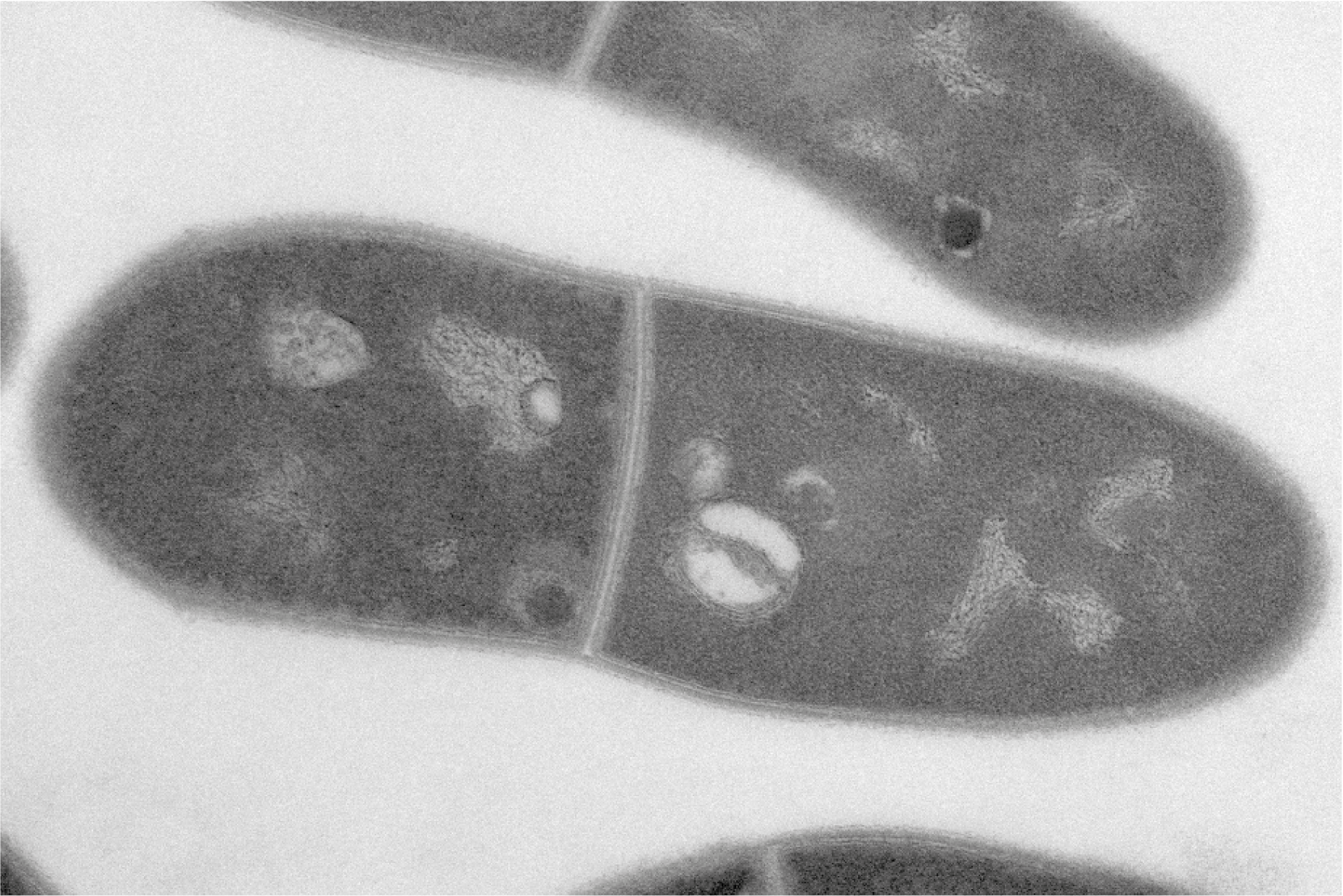

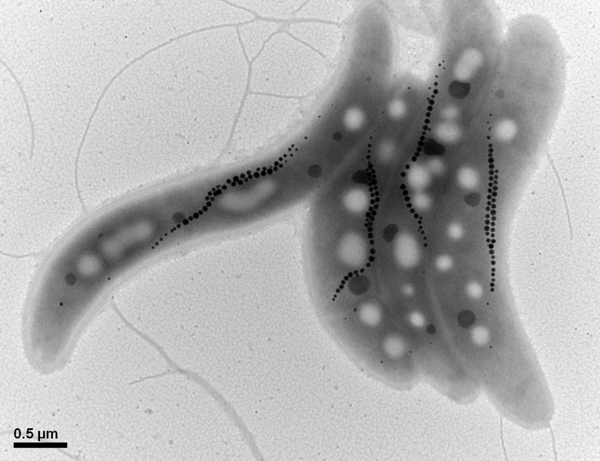
Corynebacterium glutamicum under a scanning electron microscope. The rods are not completely uniform and thus shaped like clubs (coryne in Greek). The ‘snapping’ cell division leads to unfolded V-shaped structures. Photo: Urska Repnik, Kiel University (CC BY 4.0).
Herunterladen

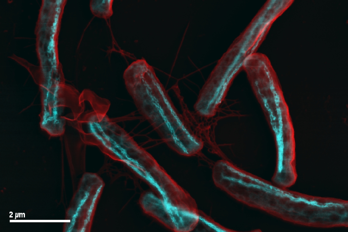
Cellular organisation of Corynebacterium glutamicum. The polar scaffold protein DivIVA shifts cell wall synthesis to the cell poles (DivIVA marked in red and cell wall synthesis in green, DNA in blue). Image: Marc Bramkamp (CC BY 4.0).
Herunterladen

Bioreactor culture of a Corynebacterium glutamicum strain developed by Metabolic Engineering for the production of astaxanthin in the feed process. Astaxanthin is an antioxidant carotenoid with an intense red colour that is used as a feed additive. Image: Florian Meyer, Bielefeld (CC BY 4.0).
Herunterladen
Press images/ files Microbe of the Year 2024 (cable bacterium Electronema)

The Microbe of the Year 2024, the cable bacterium Candidatus Electronema (magnified 10,000 times, scanning electron microscope, SEM)) forms entangled cables. Image: Pia B. Jensen, Aarhus (CC BY 4.0).
Herunterladen

Epifluorescence microscopy of Candidatus Electronema, green, FISH (EUB); blue, DAPI (Photo A. Schramm)
Herunterladen

Rice field test to stimulate cable bacteria; four measuring chambers to determine methane emissions. Source: Vincent Scholz, Tillmann Lueders.
Herunterladen

Anna Pasco Bolta: Let's symbiose and be with, performance and installation of audio-circuit cable bacteria love letter 2023. (c) Anna Pasco Bolta (http://www.annapascobolta.com/, https://www.instagram.com/apascobolta/)
Herunterladen

Overview of the different application potentials of cable bacteria, source: Scholz, Lüders
Herunterladen

Cable bacteria Candidatus Electrothrix "wires" on a slide in the laboratory (Photo: L. R. Damgaard & S. Larsen).
Herunterladen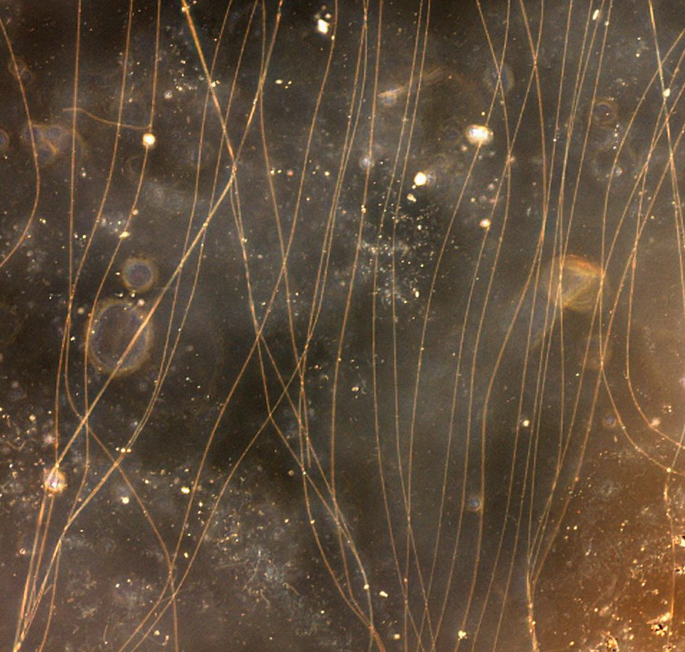

Transmission electron microscopy (TEM) of a Candidatus Electronema cross-section; the current-conducting fibres are located in the periplasm . Cell diameter 1.2 µm. Photo Pia B. Jensen, CC BY4.0
Herunterladen
Press images/ files Microbe of the Year 2023 (Bacillus subtilis)

Different lifestyles of Bacillus subtilis. Most cells express the genes
for motility (blue), only a small fraction expresses the genes for
biofilm formation (yellow), and some express none of these genes
(black). Source: Christine Diethmaier (AG Stülke), Göttingen (CC BY 4.0).

Bacillus subtilis strains can form complex biofilm structures - they
could potentially be helpful against Alzheimer's disease.
Source: Grüppen, Pelzer et al, Evonik, Halle Westfalen (CC BY 4.0).

Many people in Asia ferment soybeans with the help of Bacillus subtilis to produce traditional foods that are rich in minerals and vitamins and have a health-promoting effect, for example the Japanese Natto. Photo: Stülke, Göttingen (CC BY 4.0).
Herunterladen

Bacillus subtilis DSM 32315 heals concrete cracks. Closure of the crack by biomineralization after 92 days. Photo: Anke Reinschmidt, Interface & Performance, Evonik Operations GmbH, Essen. (CC BY 4.0)
Herunterladen

Bacillus subtilis - AG Stülke, Göttingen. (CC BY 4.0)
Herunterladen

Application of Bacillus subtilis DSM 32315 for the healing of concrete cracks. SEM image of the fracture site of a test specimen with B. subtilis spores. Photo: Anke Reinschmidt, Interface & Performance, Evonik Operations GmbH, Essen. (CC BY 4.0)
Herunterladen
Bacillus spore. Source: Pelzer, Evonik.
Herunterladen
from:
Bacillus subtilis - why this bacterium has more than deserved the title I Paulina Schneider, Dirk Schmidt I ELEMENTS - The innovation magazine by Evonik I 4/2023
Herunterladen
Press images/ files Microbe of the Year 2022 (Saccharomyces cerevisiae)

Saccharomyces cerevisiae with cell organelles. Mara Reifenrath, Frankfurt/ Main. CC BY 4.0
Herunterladen

Yeast cell (wild type), differential interference contrast. Benedikt Westermann, Bayreuth. CC BY 4.0
Herunterladen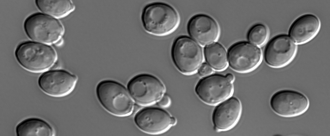

Internal structure of yeast cell with its cell organelles, electron microscope. Photo: Christina Schug, University of Bayreuth. CC BY 4.0
Herunterladen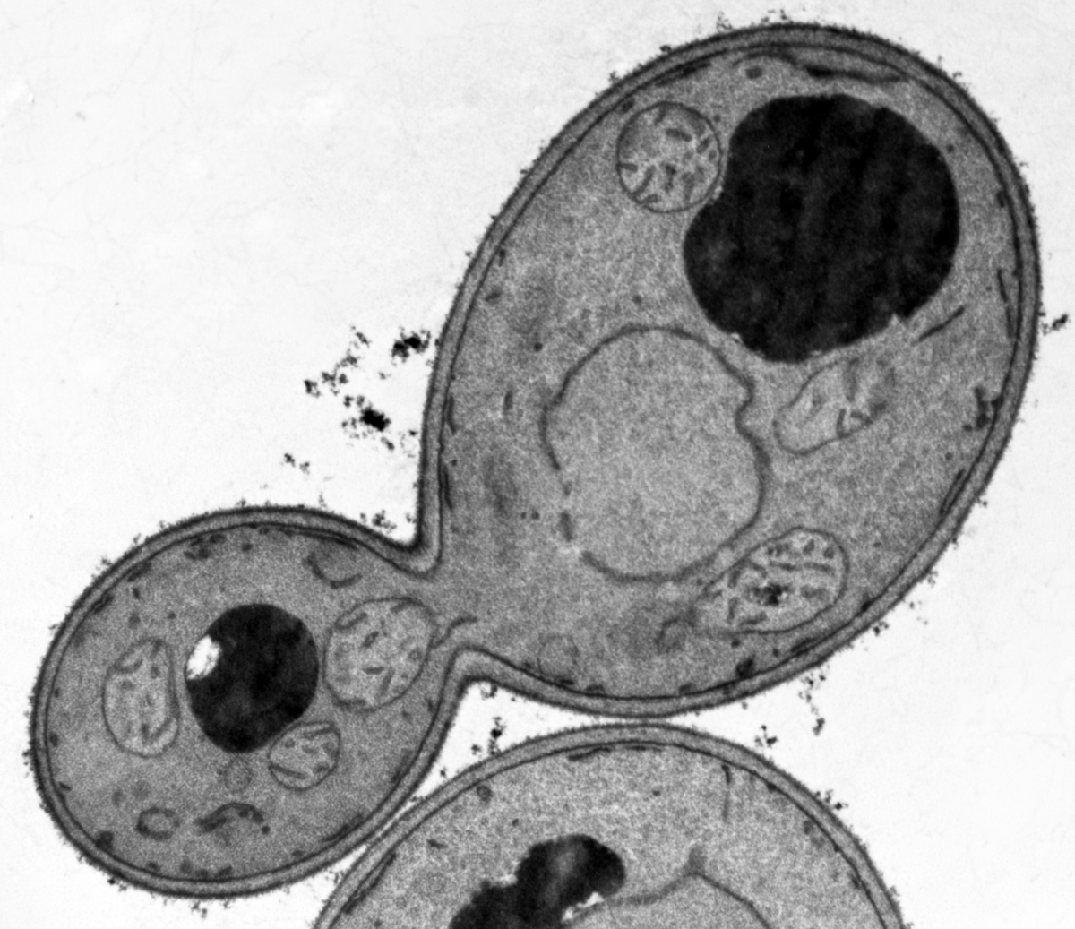

Beer made by baker´s yeast Saccharomyces cerevisiae. Photo: Benedikt Westermann, University of Bayreuth. CC BY 4.0
Herunterladen

Fermenting Futures - History of yeast Hefe from the view of art (c) Anna Dumitriu und Alex May
Herunterladen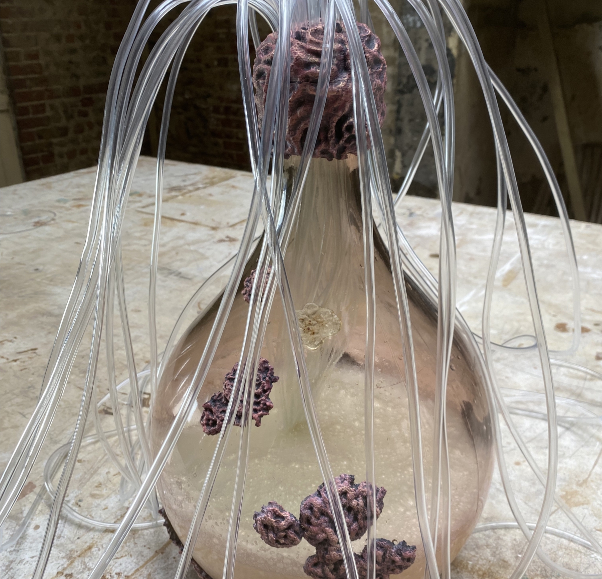

Baker´s yeast. Benedikt Westermann. CC BY 4.0
Herunterladen

Industrial plant for the production of second-generation bioethanol. In this plant, yeast produces ethanol from lignocellulose, such as straw. (c) Clariant.
Herunterladen
Pressebilder/Dateien Mikrobe des Jahres 2021 (Methanothermobacter)

Methanothermobacter thermautotrophicus im Elektronenmikroskop 30.000-fach vergrößert. (Andreas Klingl, CC BY 4.0)
Herunterladen

Methanothermobacter im Faulturm © Czichos
Herunterladen

Bioreaktor zur Erzeugung von Methan durch Methanothermobacter im Labormaßstab. Quelle: Electrochaea GmbH
Herunterladen

Pilotanlage zur Herstellung von Methangas mit Hilfe von Methanothermobacter. Quelle: Electrochaea GmbH
Herunterladen

Tauender Permafrostboden, Lena-Delta (c) www.bernhardedmaier.de
Herunterladen

Methanothermobacter. Quelle: Marco-Linus Ernst, Christian Fink, Nils Rohbohm, Tübingen
Herunterladen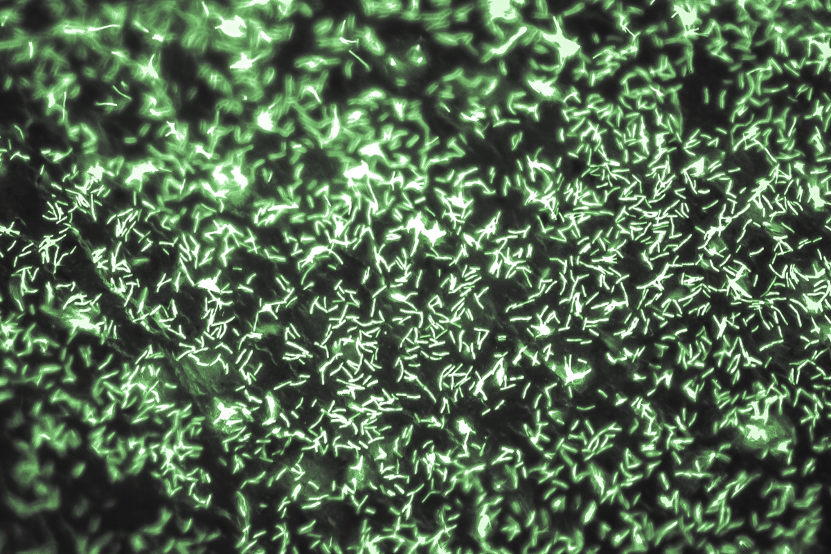

Elektrolyse erzeugt durch elektrische Energie aus Wasser Wasserstoff, den Methanothermobacter thermautotrophicus zusammen mit Kohlenstoffdioxid in Methan umwandelt (Power-to-Gas). Genetische Werkzeuge ermöglichen die Herstellung höherwertiger Produkte (Power-to-X). Die Mikroskopieaufnahme zeigt M. thermautotrophicus-Zellen, die unter dem Fluoreszenzmikroskop in 400-facher Vergrößerung mit einer Schwarz-Weiß-Kamera fotografiert und nachträglich im Computer grün eingefärbt wurden. Abb.: Bastian Molitor, Marco-Linus Ernst, Christian Fink und Nils Rohbohm, Universität Tübingen
Herunterladen

Methanothermobacter thermautotrophicus ist ein unbewegliches krummes Stäbchen. Elektronenmikroskopische Aufnahme (bearbeitet): Andreas Klingl, LMU München
Herunterladen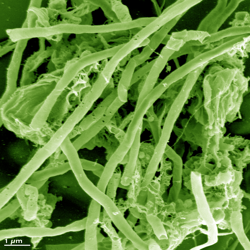
Pressebilder/Dateien Mikrobe des Jahres 2020 (Myxoxoccus xanthus)

Myxococcus xanthus jagt im Schwarm eine Escherichia coli-Kolonie. (c) Roland Garcia, HIPS
Herunterladen

M. xanthus-Zellen in Gruppen und vereinzelt (c) Treuner-Lange, MPI Marburg
Herunterladen

Hunderttausende Zellen von Myxococcus xanthus formen bei Nahrungsmangel einen kugeligen Fruchtkörper mit dauerhaften Sporen im Inneren. Quelle: http://animafactura.com/?p=100, Jürgen Berger/ Supriya Kadam, CC BY-NC 4.0
Herunterladen

Fruchtkörper von M. xanthus (c) Roland Garcia, HIPS
Herunterladen

Myxococcus xanthus-Schwarm. Foto: Ronald Garcia, HIPS (Lizensierung:CC-BY-SA-3.0)
Ein gelber Signalstoff aus Myxococcus xanthus unterstützt die Bildung von Dauerformen, die bei widrigen Bedingungen überleben können. Andere Naturstoffe aus Myxobakterien steuern das Schwarmverhalten oder wirken als Antibiotika.
Herunterladen

Video - Myxococcus xanthus-Schwarm erbeutet eine E. coli-Kolonie I HIPS (Volz)
Herunterladen
Video - Myxococcus xanthus bildet einen Fruchtkörper I HIPS (Volz)
HerunterladenMyxobacterium - Präsentation für Dozent:innen zur Mikrobe des Jahres 2020
HerunterladenWeitere Bilder finden Sie hier (Copyright beachten!)
Pressebilder/Dateien Mikrobe des Jahres 2019 (Magnetospirillum)

Pressebild Magnetospirillum
Sich teilende Zelle von Magnetospirillum gryphiswaldense mit Magnetitkristallen (Transmissions-elektronenmikroskopische Aufnahme). © Frank Mickoleit, Universität Bayreuth
Herunterladen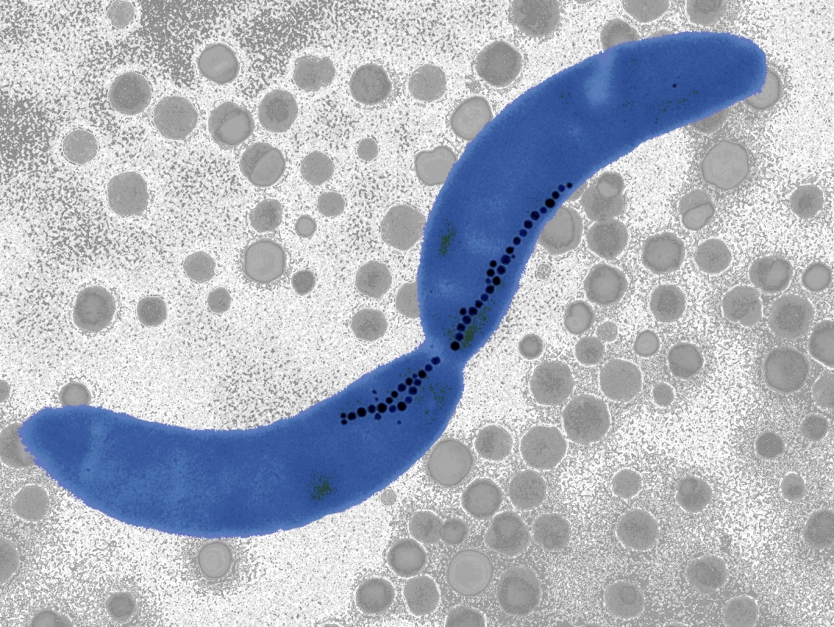

Magnetospirillum gryphiswaldense mit Magnetosomen. Foto: Frank Mickoleit/ Lizensierung: CC-BY-SA-3.0

Pressebild Magnetospirillum
Magnetospirillum gryphiswaldense in Teilung mit Magnetitkristallen (rot) und Membranvesikeln (gelb) und dem speziellen Cytoskelett (grün) sowie Geißeln zur Fortbewegung (ocker). © Mauricio Toro-Nahulepan, Universität Bayreuth/ Jürgen Plitzko, MPI für Biochemie, Martinsried
Herunterladen

Prof. Dr. Dirk Schüler, © Christian Wißler, Universität Bayreuth
Herunterladen

Pressebild Magnetospirillum
Fluoreszenz-Mikroskopie von Magnetospirillum gryphiswaldense-Zellen. Mit Grün-fluoreszierendem Protein markierte Magnetosomen-Kette, andere Zellbestandteile (Polyhydroxybutyrat-Granula) mit rotem Fluoreszenzfarbstoff angefärbt. © Frank Müller, Universität Bayreuth
Herunterladen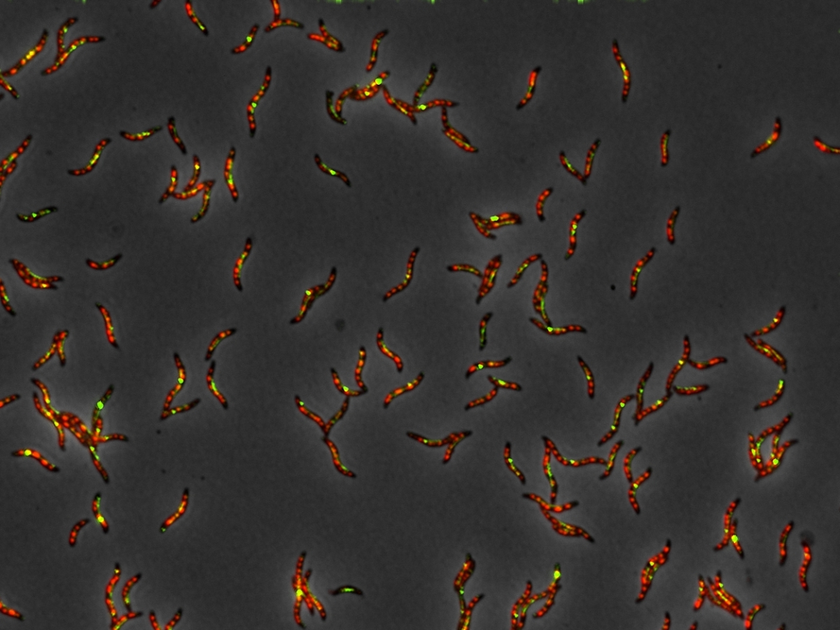


Pressebild Magnetospirillum
Rasterelektronenmikroskopische Aufnahme von M. gryphiswaldense. © Frank Müller, Universität Bayreuth
Herunterladen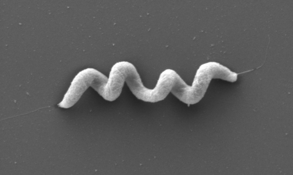

Pressebild Magnetospirillum
Magnetische vielzellige Bakterien aus dem Wattenmeer nahe Cuxhaven, von Roland Wenter et al. als Cand. Magnetomorum litorale beschrieben. © Gerhard Wanner
Herunterladen

Pressebild Magnetospirillum
Zellen von Magnetospirillum gryphiswaldense mit überaktiven Genen für die Synthese der Magnetosomen. Es bilden sich mehr als doppelt so viele und größere Magnetosomenkristalle. ©: S. Borg / A. Lohße
Herunterladen
Magnetospirillum - Präsentation für Dozenten zur Mikrobe des Jahres 2019
HerunterladenPressebilder/Dateien Mikrobe des Jahres 2018 (Lactobacillus)


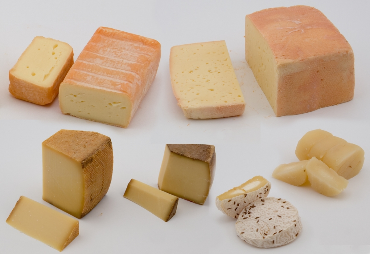
Laktobazillen sind an der Herstellung vieler Käsesorten beteiligt.
© Wilhelm Bockelmann

Lactobacillus reuteri (blau koloriert) verklumpt Helicobacter pylori (rot ) 1:11.000
© Organobalance

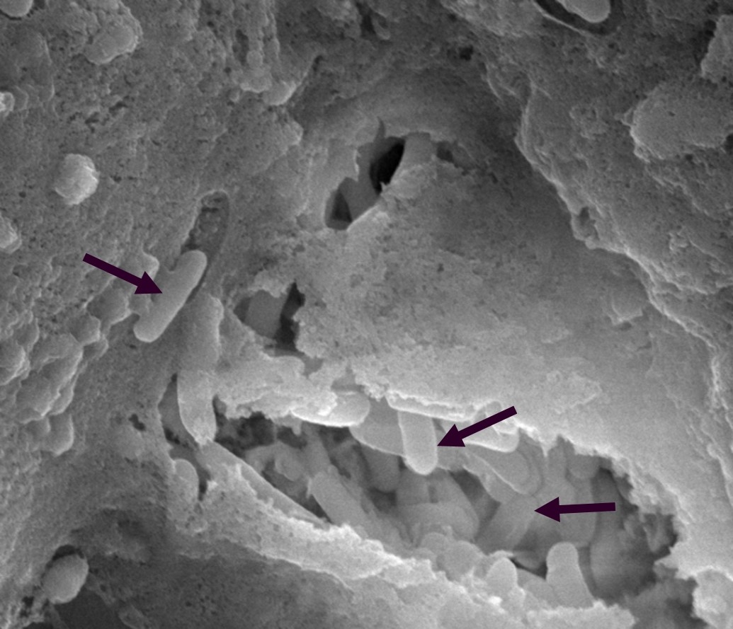
Lactobacillus salivarius (s. Pfeile) im Darm von Hühnerküken, 1:20.000
© Novozymes

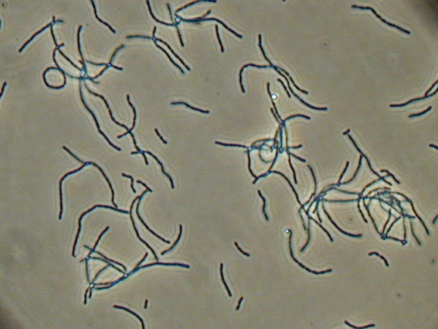
Lactobacillus rhamnosus, 1:400
© Organobalance

Lactobacillus casei, 1:400
© Organobalance

© Störiko
Herunterladen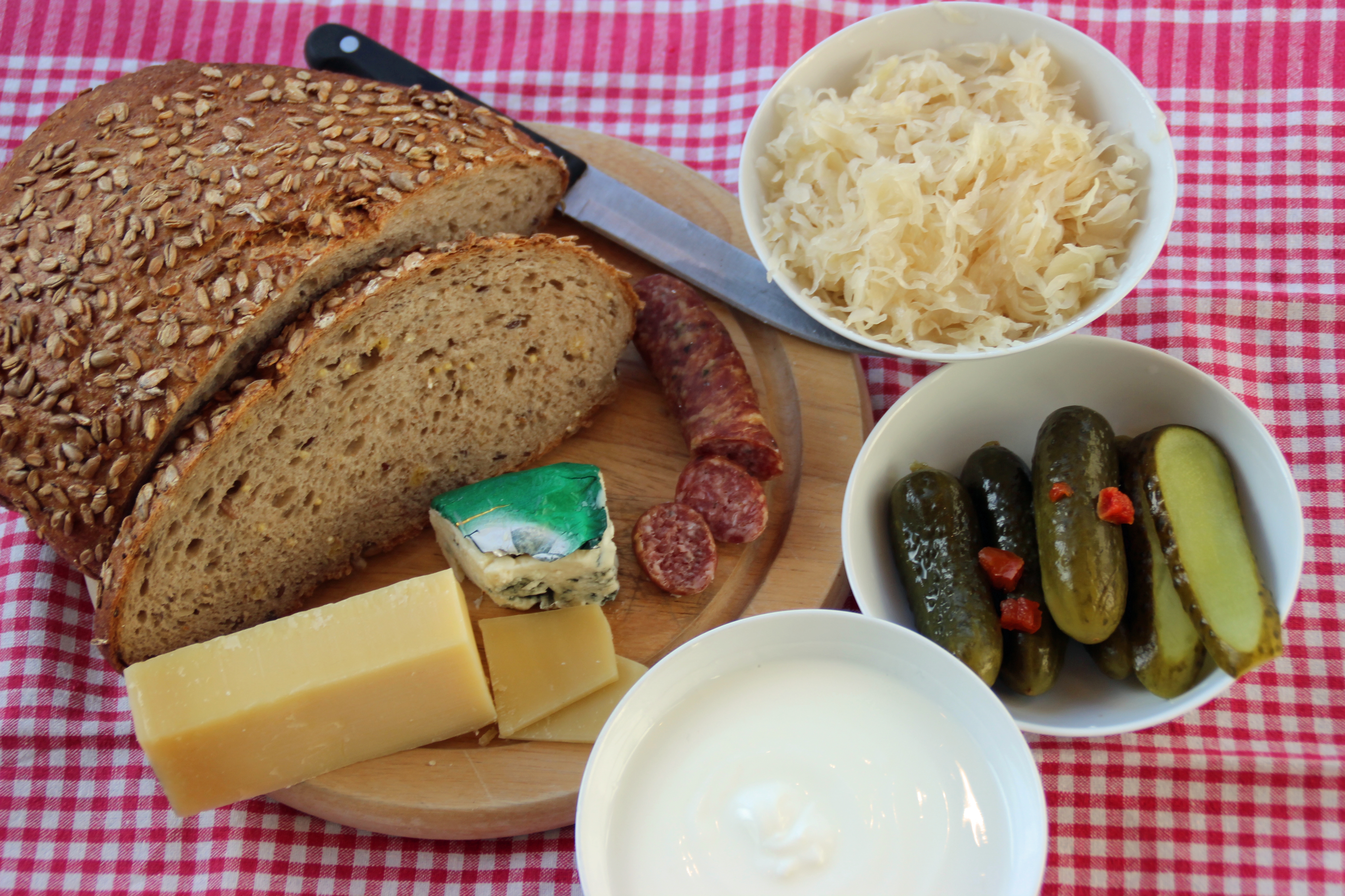

© Wikipedia/Bild-CC-by-sa/
Herunterladen

© Horst Neve, Max Rubner-Institut
Herunterladen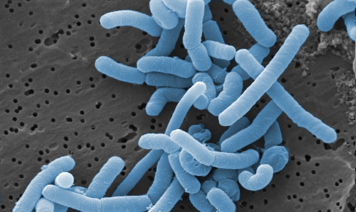

© Horst Neve, Max Rubner-Institut
Herunterladen
Pressebilder Mikrobe des Jahres 2017 (Halobacterium salinarum)

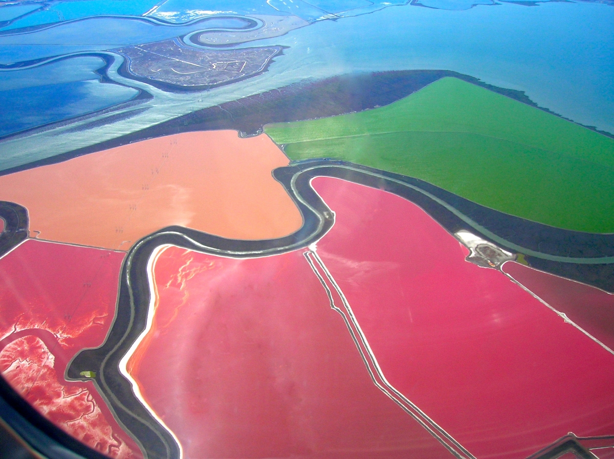
Halobacterium salinarum färben Salinen (San Francisco Bay)
Quelle: verändert nach © https://de.wikipedia.org/wiki/Saline#/media/File:San_Francisco_Bay_Salt_Ponds.jpg; Grombo, GFDL, cc-by-sa2.5,2.0,1.0

Kolonien von Halobacterium salinarum
© Felicitas Pfeifer

Isolierte Gasvesikel aus Halobacterium salinarum. Montage aus zwei Kryoelektronenmikroskopie-Aufnahmen.
© Daniel Bollschweiler, Harald Engelhardt

Bacteriorhodopsin aus Halobacterium nimmt Licht auf und verwandelt es in Energie für den Stoffwechsel der Zellen: Die Farbe des Bacteriorhodopsins wechselt dabei von violett zu gelb
© MPG, Wolfgang Filser

Die Farbstoffe aus Halobacterium salinarum färben das Gefieder von Flamingos.
© http://kostenlose-fotos.eu/Fotos/Flamingo/06-Flamingo.JPG

Salinen in Fuerteventura,
https://commons.wikimedia.org/wiki/File:
Museo_de_la_Sal,_Salinen_El_Carmen_%2810581612986%29.jpg
© Dirk Vorderstraße, Museo de la Sal
Pressebilder/Dateien Mikrobe des Jahres 2016 (Streptomyces)

Tropfen-ähnliche, Vesikel-enthaltene Exsudate einer Streptomyceten-Hyphe. Foto: Hildgund Schrempf, Osnabrück
Herunterladen

Wirkstoffe aus Streptomyceten können Bakterien abtöten – hier
sichtbar als klare Hemmhöfe im Bakterienrasen.
© Fotomontage Hildgund Schrempf, Osnabrück

Streptomyceten-Mycel mit Sporen.
© Hildgund Schrempf, Osnabrück

Streptomyceten bauen pflanzliche und tierische Reste über Zwischenstufen zu wertvoller Erde um. © Hildgund Schrempf, Osnabrück
Herunterladen

Poster zur Mikrobe des Jahres 2016 (Fotomontage: Störiko)
Herunterladen
Präsentation für Dozenten zur Mikrobe des Jahres 2016 (Streptomyces)
HerunterladenPressebilder/Dateien Mikrobe des Jahres 2015 (Rhizobium)

Bakteroidentasche im Rotklee, (c) Harald Engelhardt
Herunterladen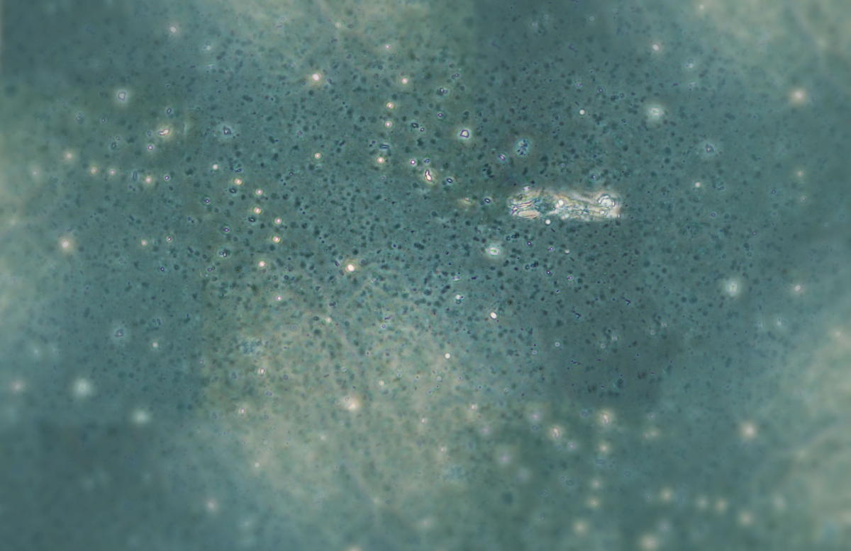

Wurzelknöllchen am Rotklee, (c) Harald Engelhardt, Martinsried
Herunterladen

Sojawurzel mit Knöllchen, rot gefärbt durch Leghämoglobin (c) Klaus-Peter Wilbois, FIBL
Herunterladen

Rotklee, (c) Harald Engelhardt
Herunterladen

Längsschnitte Sojawurzel mit Knöllchen, (c) David Kubon
Herunterladen

Detailaufnahme der Rhizobien, 1250x, (c) David Kubon
Herunterladen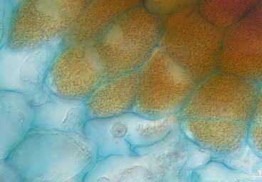

Rhizobium-Collage aus heimischen Wildkräutern (Lupine, Wicke), Nahrungsmitteln (Erbse, Bohne, Linse) und Futtermitteln (Klee), (c) Mirja Soetebier
Herunterladen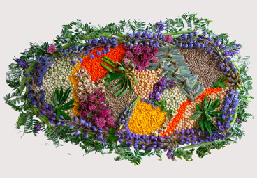
Präsentation für Dozenten zur Mikrobe des Jahres 2015
HerunterladenPressebilder/Dateien Mikrobe des Jahres 2014 (Nostoc)

Nostoc caeruleum, © Cyano Biotech GmbH, Berlin
Herunterladen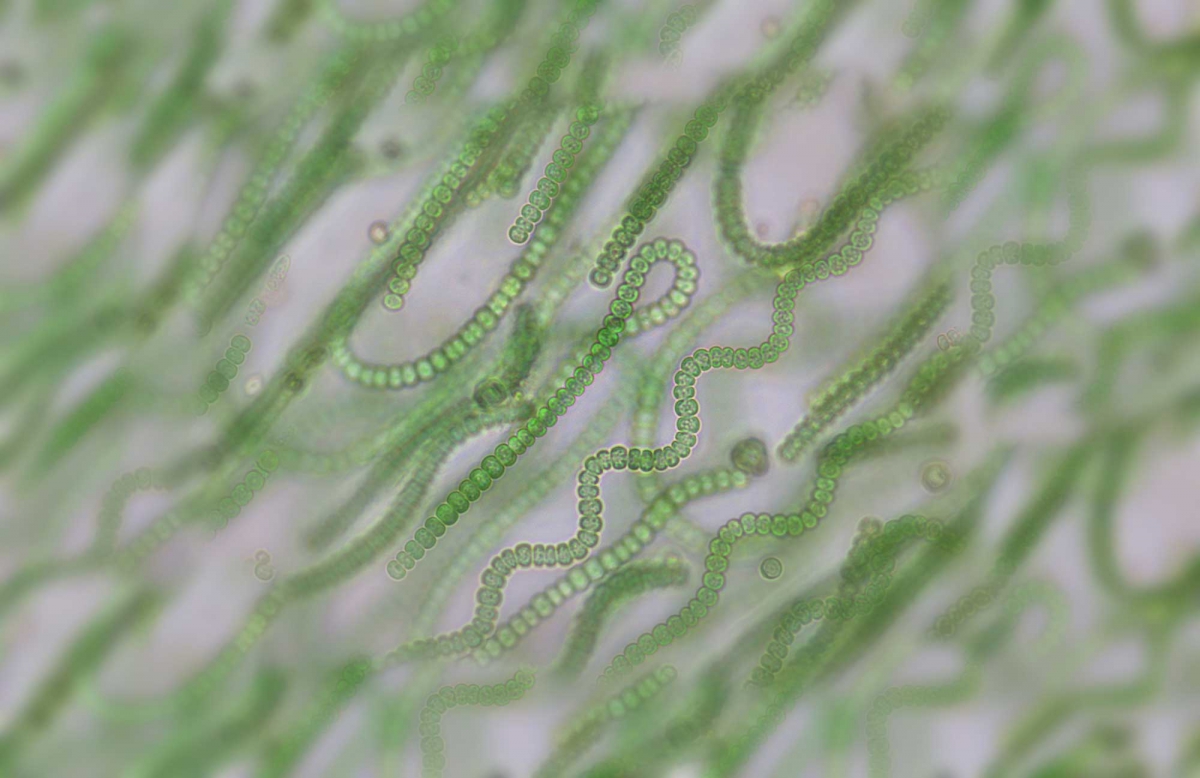

Nostoc ähnelt Pflanzen, (c) Yuuji Tsukii, Hosei University, Chiyoda-ku, Tokyo, Japan
Herunterladen

Teichpflaume Nostoc pruniforme © Christian Fischer
Herunterladen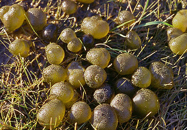

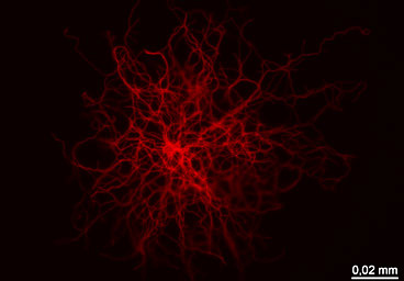
Dunkelfeld-Mikroskopie einer sphärischen Kolonie von Nostoc commune, (c) Gerd Guenther
Herunterladen

Nostoc, (c) Lena Brandt, Bad Kreuznach
Herunterladen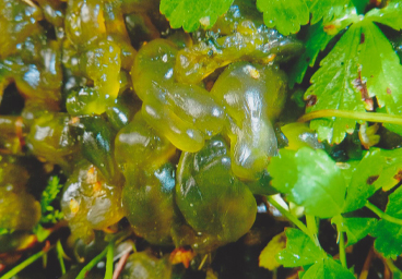
Cookie Einstellungen bearbeiten